What To Expect As You Recover from Stroke
What To Expect As You Recover from Stroke After a stroke, your life may never be the same. You will have to learn how to

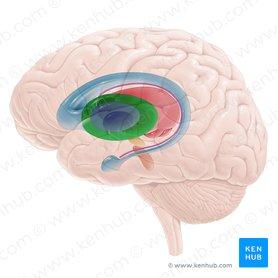
Basal ganglia
Author: Alexandru Andrușca MD, PhD • Reviewer: Dimitrios Mytilinaios MD, PhD
Last reviewed: June 30, 2022
Reading time: 27 minutes

Basal ganglia (Corpus striatum)
The basal ganglia, or basal nuclei, are a group of subcortical structures found deep within the white matter of the brain. They form a part of the extrapyramidal motor system and work in tandem with the pyramidal and limbic systems.
The basal ganglia consist of five pairs of nuclei: caudate nucleus, putamen, globus pallidus, subthalamic nucleus, and substantia nigra. These nuclei are grouped into broader clusters;
The function of the basal ganglia is to fine-tune the voluntary movements. They do so by receiving the impulses for the upcoming movement from the cerebral cortex, which they process and adjust. They convey their instructions to the thalamus, which then relays this information back to the cortex. Ultimately, the fine-tuned movement instruction is sent to the skeletal muscles through the tracts of the pyramidal motor system. Basal ganglia mediate some and other higher cortical functions as well, such as planning and modulation of movement, memory, eye movements, reward processing, and motivation.
This article will discuss the anatomy and function of the basal ganglia.
The basal ganglia are one of the components in the neural chain that controls the voluntary motor activity. The supreme component of this chain is the cerebral cortex. It generates the commands that define the motor activity of all skeletal muscles in the body. These commands descend through the pathways of the pyramidal system and synapse with the cranial nerve nuclei and motor neurons of the spinal cord. From here, the motor commands travel via the cranial and spinal nerves in order to reach the target muscles.

Basal ganglia
However, some extent of modulation and refinement of these cortical signals is necessary so that their motor execution at the muscular level happens as smoothly and precisely as planned. These adjustments are performed in the “accessory motor centers”, with the most important one being the basal ganglia. Despite being physically separated from each other, the basal ganglia are interconnected with many pathways making them a strong functional unity. Functionally, the basal ganglia are referred to as the extrapyramidal motor system although this term nowadays is not used widely. They receive and process the inputs from wide areas of the cerebral cortex, after which they relay it back to the thalamus. The thalamus then forwards those refined inputs further across the brain, mainly back to the cortex, and to the brainstem.
Phylogenetically, the oldest motor centers are the spinal cord and the reticular formation of the brainstem. With the development of the vertebrates, the brain gained new motor centers; the paleostriatum (globus pallidus) and neostriatum (caudate nucleus and putamen), which grew together with the cerebral cortex. Over time, the cerebral cortex and pyramidal system grew larger and developed a myriad of functional properties. With this, the extrapyramidal system fell under the control of the new, pyramidal, motor system, being left with the autonomy to control the nuances of cortical activity, i.e. to modulate the movements.
Recommended video: Basal ganglia
Main nuclei of the basal ganglia and surrounding structures (20 structures).
Putamen
1/7
The striatum is a complex nucleus located deep in subcortical structures of the forebrain, inside the insular lobe.
In the introduction, we mentioned that the striatum is composed of the dorsal and ventral parts. The ventral striatum is considered part of the limbic system, thus we will not describe it furthermore.
The dorsal striatum on the other hand is a component of the basal ganglia and usually, it is this part that is called “striatum” in the literature, when we describe the basal ganglia. The dorsal striatum (or simply the striatum) consists of two parts: the caudate nucleus and putamen. The parts of striatum are separated by the internal capsule, whose myelinated fibers radiate through striatum, giving it a characteristic striped appearance. Together with the globus pallidus, the striatum forms a structure called corpus striatum.
The striatum is the main input unit of the basal ganglia. It receives excitatory glutamatergic inputs from the cerebral cortex, whose synapsing pattern reflects the topography of the cortex. This means that the caudal parts of the cortex project to the caudal part of the striatum, while the rostral parts of the cortex project to the rostral part of the striatum.
The substance of the striatum is mainly (80-95%) composed of projection neurons (medium-sized spiny neurons) and minor interneurons. The projection neurons are covered by numerous spines, hence their name. Functionally, they are inhibitory neurons that use GABA as a neurotransmitter. The axons of these neurons form the direct and indirect pathways of basal ganglia, which project into the globus pallidus and substantia nigra
The interneurons of the striatum lack spines and are classified into four groups:
These neurons project to the thalamus, SNc, cerebral cortex, and control the activity of those regions.
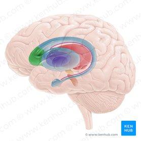
Head of caudate nucleus
Caput nuclei caudati
1/3
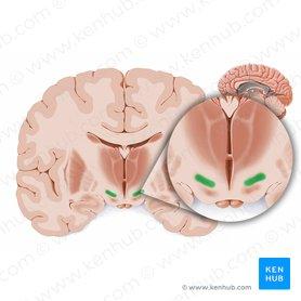
The caudate nucleus is an elongated C-shaped nucleus that lies anterior to the thalamus, just lateral to the lateral ventricles and medial to the internal capsule.
The caudate nucleus consists of the head, body and tail. The head of the nucleus contributes to the lateral wall of the lateral ventricle. The tail of the caudate nucleus forms the roof of the inferior horn of the lateral ventricle. It arches over the ventral surface of the thalamus, enters the temporal lobe and terminates by connecting with the amygdala. The rostral portion of the caudate nucleus is continuous with the putamen, and inferiorly it’s bordered by nucleus accumbens.
The functions of the caudate nucleus lie within the spectrum of functions that we described previously in the section about the striatum. More specifically, the caudate nucleus integrates sensory information about the spatial position of the body and according to that, it sends the information about the necessary fine tunes of the motor response to that stimuli to the thalamus. Additionally, it contributes to body and limb posture and the speed and accuracy of directed movements.
Besides motor control, the caudate nucleus is involved in many tasks, such as memory, goal-pursuit, learning, language processing, emotions, etc.
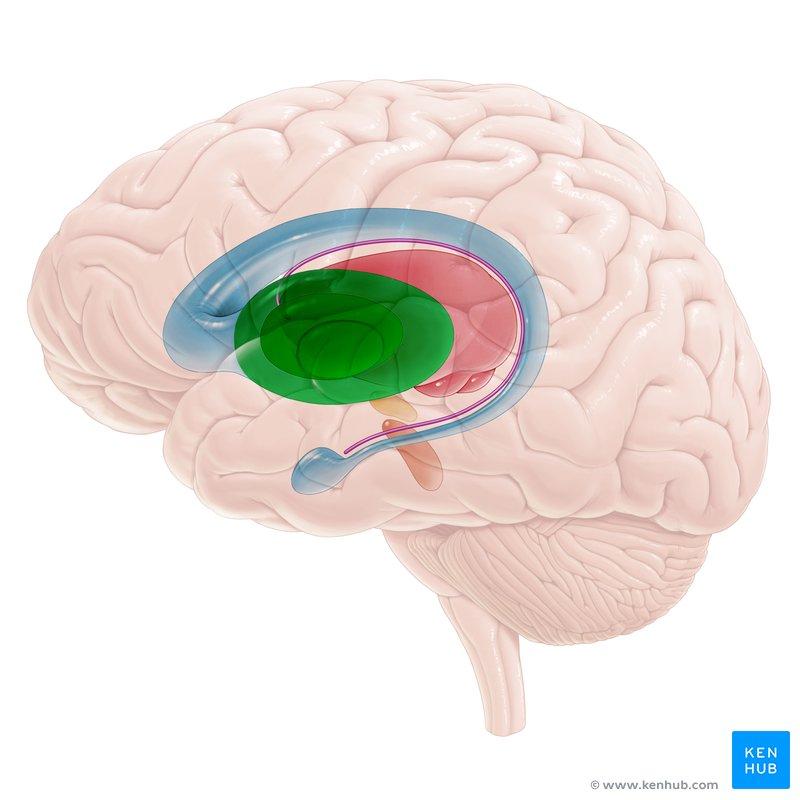
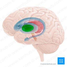
Lentiform nucleus (Nucleus lentiformis)
The putamen is a round structure situated at the base of the forebrain. It is the most lateral of the basal ganglia on the axial section of the brain. The putamen lies laterally to the globus pallidus and medially to the external capsule, covering it like a shell and extending both rostrally and caudally. It is encircled by the caudate nucleus, from which it is separated by the internal capsule.
The putamen and globus pallidus are separated by a thin layer of white matter called the medial medullary lamina.
The main function of the putamen is to regulate motor functions and influence various types of learning and it employs dopamine to perform its functions.
Nucleus accumbens and olfactory tubercle are paired structures, situated at the base of the forebrain. They are components of the ventral striatum and component of input nuclei for the ventral tegmental area (VTA).
The nucleus accumbens is found in the rostral forebrain, where the head of the caudate nucleus and putamen meet. The olfactory tubercle, however, is situated ventral to the nucleus accumbens, between the optic chiasm and olfactory tract.
Both structures are not involved in the movements regulation, rather they play an important role in the “reward circuit” and are referred to as “limbic-motor interface”. When we do anything rewarding (e.g. food, drugs, sex), dopamine neurons in an area of the brain called the ventral tegmental area (VTA) are activated. These neurons project to the nucleus accumbens and the olfactory tubercle, and when they are activated it results in an increase in dopamine levels.

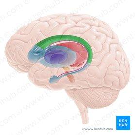
Lateral segment of globus pallidus
Globus pallidus lateralis
1/2
Synonyms: Globus pallidus lateral segment, Lateral part of globus pallidus, show more…
The globus pallidus is a paired subcortical structure, situated medially to the putamen and composed of inhibitory GABAergic projection neurons, which fire spontaneously and irregularly at high frequency. It is divided by a vertically placed sheet of white matter, the medial (internal) medullary lamina, into external (GPe) and internal (GPi) segments.
The superior and medial aspects of the globus pallidus are in contact with the internal capsule. The capsule separates the caudate nucleus from the globus pallidus. The inferior surface of the globus pallidus is in contact with the subthalamic nucleus and zona incerta, which separate it from the thalamus. Anteriorly, the globus pallidus is closely related to the substantia innominata and the hypothalamus. More caudally, it is in close proximity to the optic tract. And because the putamen and globus pallidus are in close connection, with their combined shapes resembling a bean, they are referred to as the lenticular nucleus.
Both the GPe and GPi play an essential role in the modulation of the motor program, more specifically in the direct and indirect pathways.
They both receive inhibitory GABA-ergic input from the striatum, through striatopallidal fibers, also known as Wilson’s pencils. Fibers that project from the striatum to the internal part of the globus pallidus are part of the indirect pathway of the motor loop. Meanwhile, fibers that connect the striatum with the external part of the globus pallidus are part of the direct pathway of the motor loop.
The output fibers of the globus pallidus are the pallidothalamic tracts. They divide into: ansa lenticularis, lenticular fascicles and thalamic fasciculus. Together they are part of the Forel’s field. These structures are responsible for connecting the globus pallidus and thalamic nuclei.
The globus pallidus is involved in the constant subtle regulation of movement to create smooth and precise motor actions and has a primarily inhibitory action that balances the excitatory action of the cerebellum.

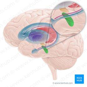
Subthalamic nucleus
Nucleus subthalamicus
1/2
Synonyms: Nucleus of Corpus Luysii, Nucleus of Luys, show more…
The subthalamic nuclei (STN), also known as Luys’ bodies, are small biconvex paired structures located within the subthalamus. The subthalamic nucleus is not an anatomical part of the basal ganglia. However, given their functional connection, the subthalamus is listed as a functional part of the basal ganglia.
The subthalamic nucleus lies at the junction of the diencephalon and midbrain, ventral to the thalamus and ventro-lateral to the red nucleus. Anteriorly its bordered by the substantia nigra and medially by the internal capsule. STN is closely related to the Forel’s fields and the pallidothalamic fibers, which entwine around its ventral and medial borders before arching back over its dorsomedial surface as the thalamic fasciculus. These fibers thus tend to separate the zone incerta from the subthalamic nucleus below and the thalamus above.
The subthalamic nuclei are composed of excitatory glutamatergic projection neurons. It receives excitatory inputs from the frontal cortex in a somatotopically organized manner. Based on this, the subthalamic nucleus is divided into three parts:
The function of the subthalamic nucleus is unknown, but some theories suggest its crucial role in the hyperdirect pathway in order to modulate the planned motor program. Additionally, considering the nucleus firing pattern, the subthalamic nucleus is considered the “pace-maker” of the basal ganglia.

Substantia nigra
1/3
Synonyms: Soemmering’s substance, Black matter, show more…
The substantia nigra is a small motor nucleus, within the anterior part of the midbrain, between the cerebral peduncle and tegmentum of the midbrain. Despite its location in the midbrain, function-wise it is considered part of basal ganglia.
The substantia nigra consists of two parts with very different connections and functions: the pars compacta (SNc) and the pars reticulata (SNr). It divides the cerebral peduncles from the tegmentum within both sides of the midbrain. Dorso-medially it is bordered by the subthalamic and red nuclei, and laterally by the medial lemniscus and the geniculate bodies.
The pars compacta comprises the dorsal portion of the substantia nigra. It consists of numerous closely packed melanin-filled neurons that give the substantia nigra its distinctive dark color. The pars reticulata lie ventral to pars compacta. It is larger than the pars compacta, but it contains fewer cells than it.
Medially to the substantia nigra is a zone called the ventral tegmental area. It is a small group of scattered cells that have similar functions to the pars compacta and may really be considered as an extension of this part.
The pars compacta serve mainly as an output to the basal ganglia circuit, supplying the striatum with dopamine, through specific D1 and D2 neurons within the nigrostriatal pathways. The pars reticulata, though, serves mainly as an input, conveying signals from the basal ganglia to the thalamus.
The loss of dopamine neurons in SNc is believed to be the reason for the development of Parkinson’s disease and some other parkinsonic syndromes.
Struggling with remembering the function of each basal nuclei? Try to understand the importance of active recall in learning anatomy.
The major efferents (outputs) of the basal ganglia consist of the neurons that project towards the thalamus and brainstem from the internal part of globus pallidus and the reticular part of the substantia nigra. These are ansa lenticularis and lenticular fasciculus.
Afferents (inputs) to the basal ganglia include the following:
In summary, the basal nuclei can be grouped functionally into four categories:
The basal nuclei modulate motor function through various pathways in order to initiate, terminate, or modulate the extent of the movement.
These are the following:


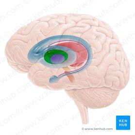
Direct pathway of the basal ganglia
1/3
Considering the conduction path and time, we can say that the hyperdirect and indirect pathways make clear initiation and termination of the selected motor program, while at the same time canceling other competing motor programs.
If you want to learn more about the pathways of the basal ganglia please read the article about the direct, indirect and hyperdirect pathways of basal ganglia.
There is a growing number of studies focused on the functions of the basal ganglia, as its functions are yet to be fully understood. However, the following are functions have been clearly established by now:
Moreover, the basal nuclei use proprioceptive feedback from the periphery to compare the movement patterns generated by the cerebral cortex with the actual movement, so that the movement is subject to ongoing refinement by a continuous servo-control mechanism.
Also, the basal ganglia have been shown to play an important role in motivation. Considering that the basal ganglia circuits are influenced heavily by extracellular dopamine, high levels of it have been linked to satiated “euphoria”, medium levels with seeking and low with aversion. The activation of the basal nuclei pathway that causes the disinhibition of the thalamus, leads to activation of the prefrontal cortex and ventral striatum. There is also evidence that other basal ganglia structures including the globus pallidus, pars medialis and subthalamic nucleus are involved in reward processing.
Regarding memory, the same structures in the prefrontal cortex are shown to be involved in the memory gates and focus. By using the basal ganglia’s direct and indirect pathways as a relay between the input information from surroundings to the cerebral structures involved in memory storage.
Now that you learned everything about the basal ganglia, test yourself and consolidate your knowledge with our quiz below!

Main nuclei of the basal ganglia and surrounding structures (20 structures).
START QUIZ
60
Basic structure identification questions
20
Advanced structure identification questions
14
Exam questions (Question bank)
Degeneration of the basal ganglia and, consequently, its dysfunction can lead to several neurological conditions. The characteristic feature of the basal ganglia lesion is a movement disorder in which there is either too little movement (hypokinesia), too much (hyperkinesia), or a combination of both, depending on the location and extent of the affected structure.
Bradykinesia represents a generalized slowness of movement and is the most common hypokinesia. The prototypical hypokinetic movement disorder is Parkinson’s disease. Parkinson’s disease results from the degeneration of the dopaminergic nigrostriatal projection. In substantia nigra pars compacta, dopaminergic neurons are decreased, so the dopaminergic output to the striatum is decreased. This leads to the reduction of the inhibition of the indirect (inhibitory) pathway and reduction of the excitation of the direct (excitatory) pathway resulting in bradykinesia, which is the main symptom of Parkinson’s disease. The condition is also characterized by resting tremor, rigidity and postural instability.
Parkinsonism is the umbrella term used to describe the symptoms of bradykinesia, tremor, and rigidity. Parkinson’s disease is the most common type of parkinsonism, but there are also some rarer types where a specific cause can be identified (ex. drug-induced parkinsonism, progressive supranuclear palsy).
The hyperkinetic movement disorders, unlike Parkinson’s disease, are characterized by too much movement. The different clinical types of hyperkinesia include dystonia, chorea, ballism, athetosis tremor, myoclonus, tics, and others.
Dystonia is characterized by involuntary, sustained muscle contraction that leads to abnormal postures of the neck, toes, hands, or other parts of the body. The exact mechanism of dystonia is not completely clear. However, the best evidence suggests that there is relevant hypoactivity in the indirect (inhibitory) pathway resulting in less inhibition and more unwanted movement. The clinical types of dystonia classify as either focal, that affects only isolated muscle groups (ex. Spasmodic Torticollis), or generalized, that typically affects muscles in the torso and limbs, and sometimes the neck and face (ex. DYT1 mutation).
Chorea, ballism, and athetosis are irregular, involuntary, jerky, and purposeless, “dance-like” movements. They are relatively similar in physiology. Ballism has a more proximal (shoulder and hip) origin and is slower than chorea. Athetosis, in nature, is slower and more twitching.
Several disorders are presenting with chorea, and the most common is Huntington’s disease. It is characterized by the degeneration of striatal GABAergic neurons, causing atrophy of the head of the caudate nucleus. Huntington’s disease is a genetic, autosomal dominant disease manifested by chorea, dementia and psychiatric abnormalities, bulbar symptoms, and gait disturbance.
Hemiballismus (ballism on the one side of the body) typically occurs after a lesion (ex. stroke, neoplasm) adjacent to the subthalamic nucleus.
Tremor is an abnormal involuntary, rhythmic and oscillatory movement of the hand, head, or other parts of the body. Usually, the basal ganglia, cerebellum, and the subthalamic nucleus are involved. However, intention tremor is also seen in disorders of the cerebellum, in which case, the tremor comes when the individual tries to perform a voluntary movement (intention tremor).
Myoclonus is a jerky, involuntary, and usually arrhythmic movement. To imagine how myoclonus looks like, think of body jerks as one is falling asleep, this is physiological myoclonus. A full list of myoclonus-related disorders is very long. Myoclonus can present in some hereditary diseases (ex. Juvenile myoclonic epilepsy) and any central nervous syndrome lesions like tumor, hemorrhage, stroke or abscess.
Tics are brief, stereotyped semi-voluntary movements, which means that unlike other movement disorders, they are partially suppressible. Tics can be either motor (motor tics) or sounds (vocal tics). They are common in children and can appear as the result of direct brain injury (ex. head trauma or encephalitis). However, most of them are idiopathic and are part of the spectrum of Gilles de la Tourette syndrome or another idiopathic tic disorder.
Most of the diseases typically have a large phenomenology of movement disorders, which includes both hypo- and hyperkinesia. In any case of young-onset parkinsonism, dystonia or other movement disorders, Wilson’s disease should be considered as this disorder is treatable and the effects of non-recognition may be severe. An autosomal recessive defect causes this disease in copper transport and the neurological manifestations are due to the accumulation of copper in basal ganglia, especially in the putamen. Because copper also accumulates in other tissues, like eyes, Kayser–Fleischer rings, a brown-green pigmentation of the Descemet membrane of the eye are diagnostic signs of Wilson’s disease.
All content published on Kenhub is reviewed by medical and anatomy experts. The information we provide is grounded on academic literature and peer-reviewed research. Kenhub does not provide medical advice. You can learn more about our content creation and review standards by reading our content quality guidelines.
References:
Illustrators:
What To Expect As You Recover from Stroke After a stroke, your life may never be the same. You will have to learn how to
How To Know If You Have A Stroke The Signs You May Be Experiencing A stroke is a serious condition that can cause paralysis on
What is a Stroke? You must have heard about stroke as a medical condition. But have you ever wondered what it means and how severe